Eye Care Technology
What's new in eye care?
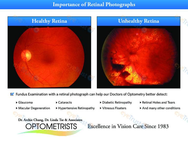
Retinal Imaging:
Retinal imaging or photography takes a digital picture of the back of your eye. It shows the retina (where light and images hit), the optic disk (a spot on the retina that holds the optic nerve, which sends information to the brain), and blood vessels. This helps your optometrist or ophthalmologist find certain diseases and check the health of your eyes.
Optometrist have long used a tool called an ophthalmoscope to look at the back of your eye. Retinal imaging allows Optometrist to get a much wider digital view of the retina and adds a more detailed assessment during your comprehensive eye health exam.
Currently, there are two main types of retinal imaging. The first type of retinal imaging is using standard equipment that can image 45º of the retina. The second type of retinal imaging is using advanced equipment that can image 220º of the retina. Ask your Optometrist or eye care professional about optos technology.
Who Gets This Test?
Your Optometrist may recommend retinal imaging if you have any of the following diseases or conditions:
Diabetes: This disease can damage the blood vessels in your retina. Over time, it causes you to lose your sight if it is not controlled.
Macular degeneration : The central part of your retina (the macula) starts to get worse with age. You may have blurry vision and find it harder to focus. If that happens, you may be considered legally blind even though you may still have peripheral vision. There are two kinds of macular degeneration: wet and dry.
Dry macular degeneration is by far the most common form of this disease (up to 90% of the cases). It happens when blood vessels under the retina become thin and brittle. Abnormal blood vessels growing under the retina cause wet macular degeneration. Vision loss is usually fast. Retinal imaging is very important in finding this type of macular degeneration.
Glaucoma : This disease damages your optic nerve (located in the retina) and may cause vision loss. It typically happens when fluid builds up in the front of your eye. It can cause blindness but it normally progresses slowly and can be treated with special eye drops to lower the pressure caused by the fluid.
Visit your Optometrist or eye care professional to learn more about retinal imaging and the benefits of early detection for eye conditions and diseases.
Optical Coherence Tomography (OCT)
Optical coherence tomography (OCT) is a non-invasive imaging test. OCT uses light waves to take cross-section pictures of your retina.
With OCT, your Optometrist or Ophthalmologist can see each of the retina’s distinctive layers. This allows your Optometrist or Ophthalmologist to map and measure their thickness. These measurements help with diagnosis as they provide treatment guidance for glaucoma and diseases of the retina. These retinal diseases include age-related macular degeneration (AMD) and diabetic eye disease.
Visual Field Analyzer:
Visual field analyzers are often used in detection and diagnosis of Glaucoma, AMD, Scotomas and brain abnormalities. These analyzers test the full horizontal and vertical field of peripheral vision, identifying potential diminished or blind spots (scotomas) in the patients’ vision. Identifying specific scotomas in ones vision can be indicative of a certain type of brain trauma or disease, such as a stroke or tumour. The patient is directed to identify certain light stimuli provided by the analyzer in their peripheral vision, if the patient is unable to detect the stimuli, then this could indicate trauma and/or scotomas.
These visual field analyzers can offer up to 90º of measurement across the visual field and boast features such as monocular and binocular testing, fast and simple operation, colour and kinetic perimetry to name just a few. To learn more about visual field testing, click here.
Corneal Topography
Corneal topography is a computerized test that maps the curve of your cornea. It can show problems with your eye’s surface, like swelling or scarring, or conditions such as astigmatism. You might have it before you get surgery, a cornea transplant, or a contact lens fitting.